Diagnosis and Treatment Protocol for Novel Coronavirus Pneumonia (Trial Version 7)

(Released by National Health Commission & State Administration of Traditional Chinese Medicine on March 3, 2020)
Since December 2019, multiple cases of novel coronavirus pneumonia (NCP) have been identified in Wuhan, Hubei. With the spread of the epidemic, such cases have also been found in other parts of China and other countries. By taking a series of preventive control and medical treatment measures, the rise of the epidemic situation in China has been contained to a certain extent, and the epidemic situation has eased in most provinces, but the incidence abroad is on the rise. With increased understanding of the clinical manifestations and pathology of the disease, and the accumulation of experience in diagnosis and treatment, in order to further strengthen the early diagnosis and early treatment of the disease, improve the cure rate, reduce the mortality rate, avoid nosocomial infection as much as possible and pay attention to the spread caused by the imported cases from overseas, we revised the Diagnosis and Treatment Protocol for Novel Coronavirus Pneumonia (Trial Version 6) to Diagnosis and Treatment Protocol for Novel Coronavirus Pneumonia (Trial Version 7).
I .Etiological Characteristics
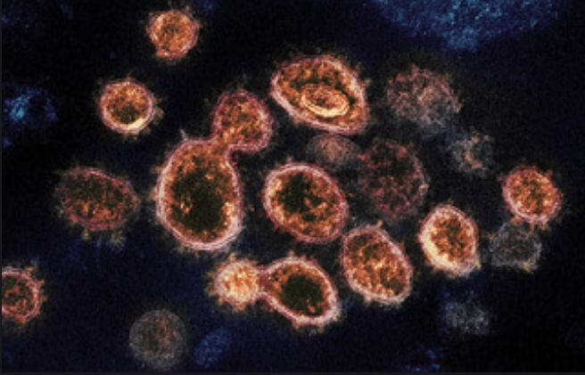
The novel coronaviruses belong to the β genus. They have envelopes, and the particles are round or oval, often polymorphic, with diameter being 60 to 140 nm. Their genetic characteristics are significantly different from SARS-CoV and MERS-CoV. Current research shows that they share more than 85% homology with bat SARS-like coronaviruses (bat-SL-CoVZC45). When isolated and cultured in vitro, the 2019-nCoV can be found in human respiratory epithelial cells in about 96 hours, however it takes about 6 days for the virus to be found if isolated and cultured in Vero E6 and Huh-7 cell lines.
Most of the know-how about the physical and chemical properties of coronavirus comes from the research on SARS-CoV and MERS-CoV. The virus is sensitive to ultraviolet and heat. Exposure to 56°C for 30 minutes and lipid solvents such as ether, 75% ethanol, chlorine-containing disinfectant, peracetic acid, and chloroform can effectively inactivate the virus. Chlorhexidine has not been effective in inactivating the virus.
II. Epidemiological Characteristics
1.Source of infection
Now, the patients infected by the novel coronavirus are the main source of infection; asymptomatic infected people can also be an infectious source.
2.Route of transmission
Transmission of the virus happens mainly through respiratory droplets and close contact. There is the possibility of aerosol transmission in a relatively closed environment for a long-time exposure to high concentrations of aerosol. As the novel coronavirus can be isolated in feces and urine, attention should be paid to feces or urine contaminated environmental that leads to aerosol or contact transmission.
3.Susceptible groups
People are generally susceptible.
III. Pathological changes
Pathological findings from limited autopsies and biopsy studies.
1.Lungs
Solid changes of varying degrees are present in the lungs. Alveolar damage involves fibromyxoid exudation and hyaline membrane formation. The exudates are composed of monocytes and macrophages, with plenty of multinucleated syncytial cells. Type II alveolar epithelial cells are markedly hyperplastic, some of which are desquamated. Viral inclusions are observed in type II alveolar epithelial cells and macrophages. Alveolar interstitium is marked with vascular congestion and edema, infiltration of monocytes and lymphocytes, and vascular hyaline thrombi. The lungs are laden with hemorrhagic and necrotic foci, along with evidence of hemorrhagic infarction. Organization of alveolar exudates and interstitial fibrosis are partly present.
The bronchi are filled with desquamated epithelial cells, mucus and mucus plugs. Hyperventilated alveoli, interrupted alveolar interstitium and cystic formation are occasionally seen.
2.Spleen, hilar lymph nodes and bone marrow
The spleen is evidently shrunk. Lymphocytopenia and focal hemorrhage and necrosis are present. Macrophagocyte proliferation and phagocytosis are noted in the spleen. Lymph nodes are found with sparse lymphocytes and occasional necrosis. CD4+ and CD8+ T cells are present in reduced quantity in the spleen and lymph nodes, revealed by immunohistochemistry staining. Pancytopenia is identified in bone marrow.
3.Heart and blood vessels
Degenerated or necrosed myocardial cells are present, along with mild infiltration of monocytes, lymphocytes and/or neutrophils in the cardiac interstitium. Endothelial desquamation, endovasculitis and thrombi are seen in some blood vessels.
4.Liver and gall bladder
Appearing enlarged and dark-red, the liver is found degenerated with focal necrosis infiltrated with neutrophils. The liver sinusoids are found hyperemic. The portal areas are infiltrated with lymphocytes and monocytes and dotted with microthrombi. The gall bladder is prominently filled.
5.Kidneys
The kidneys are noted with protein exudation in the Bowman’s capsule around glomeruli, degeneration and desquamation of the epithelial cells of renal tubules, and hyaline casts. Microthrombi and fibrotic foci are found in the kidney interstitium.
6.Other organs
Cerebral hyperemia and edema are present, with degeneration of some neurons. Necrosis foci are noted in the adrenal glands. Degeneration, necrosis and desquamation of epithelium mucosae at varying degrees are present in the esophageal, stomach and intestine.
IV. Clinical Characteristics
1.Clinical manifestations
Based on the current epidemiological investigation, the incubation period is one to 14 days, mostly three to seven days.
Main manifestations include fever, fatigue and dry cough. Nasal congestion, runny nose, sore throat, myalgia and diarrhea are found in a few cases. Severe cases mostly developed dyspnea and/or hypoxemia after one week. In severe cases, patients progress rapidly to acute respiratory distress syndrome, septic shock, metabolic acidosis that is difficult to correct, coagulopathy, multiple organ failure and others. It is worth noting that for severe and critically ill patients, their fever could be moderate to low, or even barely noticeable. Some children and neonatal cases may have atypical symptoms, manifested as gastrointestinal symptoms such as vomiting and diarrhea, or only manifested as low spirits and shortness of breath. The patients with mild symptoms did not develop pneumonia but only low fever and mild fatigue. From current situations, most patients have good prognosis and a small number of patients are critically ill. The prognosis for the elderly and patients with chronic underlying diseases is poor. The clinical course of pregnant women with NCP is similar to that of patients of the same age. Symptoms in children are relatively mild.
2.Laboratory tests
General findings
In the early stages of the disease, peripheral WBC count is normal or decreased and the lymphocyte count decreases. Some patients see an increase in liver enzymes, lactate dehydrogenase (LDH), muscle enzymes and myoglobin. Elevated troponin is seen in some critically ill patients while most patients have elevated C-reactive protein and erythrocyte sedimentation rate and normal procalcitonin. In severe cases, D-dimer increases and peripheral blood lymphocytes progressively decrease. Severe and critically ill patients often have elevated inflammatory factors.
Pathogenic and serological findings
(1) Pathogenic findings: Novel coronavirus nucleic acid can be detected in nasopharyngeal swabs, sputum, lower respiratory tract secretions, blood, feces and other specimens using RT-PCR and/or NGS methods. It is more accurate if specimens from lower respiratory tract (sputum or air tract extraction) are tested. The specimens should be submitted for testing as soon as possible after collection.
(2) Serological findings: NCP virus specific IgM becomes detectable around 3-5 days after onset; IgG reaches a titration of at least 4-fold increase during convalescence compared with the acute phase.
3.Chest imaging
In the early stage, imaging shows multiple small patchy shadows and interstitial changes, apparent in the outer lateral zone of lungs. As the disease progresses, imaging then shows multiple ground glass opacities and infiltration in both lungs. In severe cases, pulmonary consolidation may occur while pleural effusion is rare.
V. Case Definitions
1.Suspect cases
Considering both the following epidemiological history and clinical manifestations:
1.1 Epidemiological history
1.1.1 History of travel to or residence in Wuhan and its surrounding areas, or in other communities where cases have been reported within 14 days prior to the onset of the disease;
1.1.2 In contact with novel coronavirus infected people (with positive results for the nucleic acid test) within 14 days prior to the onset of the disease;
1.1.3 In contact with patients who have fever or respiratory symptoms from Wuhan and its surrounding area, or from communities where confirmed cases have been reported within 14 days before the onset of the disease; or
1.1.4 Clustered cases (2 or more cases with fever and/or respiratory symptoms in a small area such families, offices, schools etc within 2 weeks). 1.2 Clinical manifestations
1.2.1 Fever and/or respiratory symptoms;
1.2.2 The aforementioned imaging characteristics of NCP;
1.2.3 Normal or decreased WBC count, normal or decreased lymphocyte count in the early stage of onset.
A suspect case has any of the epidemiological history plus any two clinical manifestations or all three clinical manifestations if there is no clear epidemiological history.
2.Confirmed cases
Suspect cases with one of the following etiological or serological evidences:
2.1 Real-time fluorescent RT-PCR indicates positive for new coronavirus nucleic acid; 2.2 Viral gene sequence is highly homologous to known new coronaviruses.
2.3 NCP virus specific Ig M and IgG are detectable in serum; NCP virus specific IgG is detectable or reaches a titration of at least 4-fold increase during convalescence compared with the acute phase.
VI. Clinical Classification
1.Mild cases
The clinical symptoms were mild, and there was no sign of pneumonia on imaging.
2. Moderate cases
Showing fever and respiratory symptoms with radiological findings of pneumonia.
3. Severe cases
Adult cases meeting any of the following criteria:
(1) Respiratory distress (≧30 breaths/ min);
(2) Oxygen saturation≤93% at rest;
(3) Arterial partial pressure of oxygen (PaO2)/ fraction of inspired oxygen (FiO2)≦
300mmHg (l mmHg=0.133kPa).
In high-altitude areas (at an altitude of over 1,000 meters above the sea level), PaO2/ FiO2 shall be corrected by the following formula:
PaO2/ FiO2 x[Atmospheric pressure (mmHg)/760]
Cases with chest imaging that showed obvious lesion progression within 24-48 hours >50% shall be managed as severe cases.
Child cases meeting any of the following criteria:
(1) Tachypnea (RR ≥ 60 breaths/min for infants aged below 2 months; RR ≥ 50 BPM for infants aged 2-12 months; RR ≥ 40 BPM for children aged 1-5 years, and RR ≥ 30 BPM for children above 5 years old) independent of fever and crying;
(2) Oxygen saturation ≤ 92% on finger pulse oximeter taken at rest;
(3) Labored breathing (moaning, nasal fluttering, and infrasternal, supraclavicular and intercostal retraction), cyanosis, and intermittent apnea;
(4) Lethargy and convulsion;
(5) Difficulty feeding and signs of dehydration.
4.Critical cases
Cases meeting any of the following criteria:
4.1 Respiratory failure and requiring mechanical ventilation; 4.2 Shock;
4.3 With other organ failure that requires ICU care.
VII. Clinical early warning indicators of severe and critical cases
1.Adults. 1.1 The peripheral blood lymphocytes decrease progressively; 1.2 Peripheral blood inflammatory factors, such as IL-6 and C-reactive proteins, increase progressively;
1.3 Lactate increases progressively;
1.4 Lung lesions develop rapidly in a short period of time.
2.Children.
2.1 Respiratory rate increased;
2.2 Poor mental reaction and drowsiness;
2.3 Lactate increases progressively;
2.4 Imaging shows infiltration on both sides or multiple lobes, pleural effusion or rapid progress of lesions in a short period of time;
2.5 Infants under the age of 3 months who have either underlying diseases (congenital heart disease, bronchopulmonary dysplasia, respiratory tract deformity, abnormal hemoglobin, and severe malnutrition, etc.) or immune deficiency or hypofunction (long-term use of immunosuppressants).
VIII. Differential Diagnosis
The mild manifestations of NCP need to be distinguished from upper respiratory tract infections caused by other viruses.
NCP is mainly distinguished from other known viral pneumonia and mycoplasma pneumoniae infections such as influenza virus, adenovirus and respiratory syncytial virus. Especially for suspect cases, methods such as rapid antigen detection and multiplex PCR nucleic acid detection should be adopted as much as possible for detection of common respiratory pathogens.
It should also be distinguished from non-infectious diseases such as vasculitis, dermatomyositis and organizing pneumonia.
IX. Case Finding and Reporting
Health professionals in medical institutions of all types and at all levels, upon discovering suspect cases that meet the definition, should immediately put them in single room for isolation and treatment. If the cases are still considered as suspected after consultation made by hospital experts or attending physicians, it should be reported directly online within 2 hours; samples should be collected for new coronavirus nucleic acid testing and suspect cases should be safely transferred to the designated hospitals immediately. People who have been in close contact with patients who have been confirmed of new coronavirus infection are advised to perform new coronavirus pathogenic testing in a timely manner, even though common respiratory pathogens are tested positive.
If two nucleic acid tests, taken at least 24-hour apart, of a NCP suspect case are negative, and the NCP virus specific IgM and IgG are negative after 7 days from onset, the suspect diagnosis can be ruled out.
X. Treatment
1.Treatment venue determined by the severity of the disease
1.1 Suspected and confirmed cases should be isolated and treated at designated hospitals with effective isolation, protection and prevention conditions in place. A suspect case should be treated in isolation in a single room. Confirmed cases can be treated in the same room.
1.2 Critical cases should be admitted to ICU as soon as possible.
2.General treatment
2.1 Letting patients rest in bed and strengthening support therapy; ensuring sufficient caloric intake for patients; monitoring their water and electrolyte balance to maintain internal environment stability; closely monitoring vital signs and oxygen saturation.
2.2 According to patients’ conditions, monitoring blood routine result, urine routine result, c-reactive protein (CRP), biochemical indicators (liver enzyme, myocardial enzyme, renal function etc.), coagulation function, arterial blood gas analysis, chest imaging and cytokines detection if necessary.
2.3 Timely providing effective oxygen therapy, including nasal catheter and mask oxygenation and nasal high-flow oxygen therapy. If possible, inhalation of mixed hydrogen and oxygen (H2/O2: 66.6%/33.3%) can be applied.
2.4 Antiviral therapy: Hospitals can try Alpha-interferon (5 million U or equivalent dose each time for adults, adding 2ml of sterilized water, atomization inhalation twice daily), lopinavir/ritonavir (200 mg/50mg per pill for adults, two pills each time, twice daily, no longer than 10 days), Ribavirin (suggested to be used jointly with interferon or lopinavir/ritonavir, 500 mg each time for adults, twice or three times of intravenous injection daily, no longer than 10 days), chloroquine phosphate (500 mg bid for 7 days for adults aged 18-65 with body weight over 50 kg; 500 mg bid for Days 1&2 and 500 mg qd for Days 3-7 for adults with body weight below 50 kg), Arbidol (200 mg tid for adults, no longer than 10 days). Be aware of the adverse reactions, contraindications (for example, chloroquine cannot be used for patients with heart diseases) and interactions of the above- mentioned drugs. Further evaluate the efficacy of those drugs currently being used. Using three or more antiviral drugs at the same time is not recommend; if an intolerable toxic side effect occurs, the respective drug should be discontinued. For the treatment of pregnant women, issues such as the number of gestational weeks, choice of drugs having the least impact on the fetus, as well as whether pregnancy being terminated before treatment should be considered with patients being informed of these considerations.
2.5 Antibiotic drug treatment: Blind or inappropriate use of antibiotic drugs should be avoided, especially in combination with broad-spectrum antibiotics.
3.Treatment of severe and critical cases
3.1 Treatment principle: On the basis of symptomatic treatment, complications should be proactively prevented, underlying diseases should be treated, secondary infections also be prevented, and organ function support should be provided timely.
3.2 Respiratory support:
3.2.1 Oxygen therapy: Patients with severe symptoms should receive nasal cannulas or masks for oxygen inhalation and timely assessment of respiratory distress and/or hypoxemia should be performed.
3.2.2 High-flow nasal-catheter oxygenation or noninvasive mechanical ventilation: When respiratory distress and/or hypoxemia of the patient cannot be alleviated after receiving standard oxygen therapy, high-flow nasal cannula oxygen therapy or non-invasive ventilation can be considered. If conditions do not improve or even get worse within a short time (1-2 hours), tracheal intubation and invasive mechanical ventilation should be used in a timely manner.
3.2.3 Invasive mechanical ventilation: Lung protective ventilation strategy, namely low tidal volume (6-8ml/kg of ideal body weight) and low level of airway platform pressure (<30cmH2O) should be used to perform mechanical ventilation to reduce ventilator-related lung injury. While the airway platform pressure maintained ≤30cmH2O, high PEEP can be used to keep the airway warm and moist; avoid long sedation and wake the patient early for lung rehabilitation. There are many cases of human-machine asynchronization, therefore sedation and muscle relaxants should be used in a timely manner. Use closed sputum suction according to the airway secretion, if necessary, administer appropriate treatment based on bronchoscopy findings.
3.2.4 Rescue therapy: Pulmonary re-tensioning is recommended for patients with severe ARDS. With sufficient human resources, prone position ventilation should be performed for more than 12 hours per day. If the outcome of prone position ventilation is poor, extracorporeal membrane oxygenation (ECMO) should be considered as soon as possible. Indications include: 1When Fi02>90%, the oxygenation index is less than 80mmHg for
more than 3-4 hours; 2For patients with only respiratory failure when the airway platform pressure ≥ 35cmH2O, VV-ECMO mode is preferred; if circulatory support is needed, VA- ECMO mode should be used. When underlying diseases are under control and the cardiopulmonary function shows signs of recovery, withdrawal of ECMO can be tried. 3.3 Circulatory support: On the basis of adequate fluid resuscitation, efforts should be made to improve microcirculation, use vasoactive drugs, closely monitor changes in blood pressure, heart rate and urine volume as well as lactate and base excess in arterial blood gas analysis. If necessary, use non-invasive or invasive hemodynamic monitor such as Doppler ultrasound, echocardiography, invasive blood pressure or continuous cardiac output (PiCCO) monitoring. In the process of treatment, pay attention to the liquid balance strategy to avoid excessive or insufficient fluid intake.
If the heart rate suddenly increases more than 20% of the basic value or the decrease of blood pressure is more than 20% of the basic value with manifestations of poor skin perfusion and decreased urine volume, make sure to closely observe whether the patient has septic shock, gastrointestinal hemorrhage or heart failure.
3.4 Renal failure and renal replacement therapy: Active efforts should be made to look for causes for renal function damage in critical cases such as low perfusion and drugs. For the treatment of patients with renal failure, focus should be on the balance of body fluid, acid and base and electrolyte balance, as well as on nutrition support including nitrogen balance and the supplementation of energies and trace elements. For critical cases, continuous renal replacement therapy (CRRT) can be used. The indications include: 1 hyperkalemia; 2 acidosis; 3 pulmonary edema or water overload; 4 fluid management in multiple organ dysfunction.
3.5 Convalescent plasma treatment: It is suitable for patients with rapid disease progression, severe and critically ill patients. Usage and dosage should refer to Protocol of Clinical Treatment with Convalescent Plasma for NCP Patients (2nd trial version).
3.6 Blood purification treatment: Blood purification system including plasma exchange, absorption, perfusion and blood/plasma filtration can remove inflammatory factors and block “cytokine storm”, so as to reduce the damage of inflammatory reactions to the body. It can be used for the treatment of severe and critical cases in the early and middle stages of cytokine storm.
3.7 Immunotherapy: For patients with extensive lung lesions and severe cases who also show an increased level of IL-6 in laboratory testing, Tocilizumab can be used for treatment. The initial dose is 4-8mg/kg with the recommended dose of 400mg diluted with 0.9% normal saline to 100ml. The infusion time should be more than 1 hour. If the initial medication is not effective, one extra administration can be given after 12 hours (same dose as before). No more than two administrations should be given with the maximum single dose no more than 800mg. Watch out for allergic reactions. Administration is forbidden for people with active infections such as tuberculosis.
3.8 Other therapeutic measures
For patients with progressive deterioration of oxygenation indicators, rapid progress in imaging and excessive activation of the body’s inflammatory response, glucocorticoids can be used in a short period of time (three to five days). It is recommended that dose should not exceed the equivalent of methylprednisolone 1-2 mg/kg/day. Note that a larger dose of glucocorticoid will delay the removal of coronavirus due to immunosuppressive effects. Xuebijing 100ml/time can be administered intravenously twice a day. Intestinal microecological regulators can be used to maintain intestinal microecological balance and prevent secondary bacterial infections.
Child severe and critical cases can be given intravenous infusion of γ-globulin.
For pregnant severe and critical cases, pregnancy should be terminated preferably with c- section.
Patients often suffer from anxiety and fear and they should be supported by psychological counseling.
4.Traditional Chinese Medicine treatment
Goodluck to everyone.